ANNELID LAB
PURPOSE: To compare and contrast the internal and external body parts of annelids.
MATERIALS:
- Preserved earthworm
- Magnifying glass or hand lens
- Dissection tray
- Pins
- Dissection kit
- Gloves
- Microscope and slides
PURPOSE: To compare and contrast the internal and external body parts of annelids.
MATERIALS:
- Preserved earthworm
- Magnifying glass or hand lens
- Dissection tray
- Pins
- Dissection kit
- Gloves
- Microscope and slides
|
METHOD: EARTHWORM (OLIGOCHAETA) DISSECTION
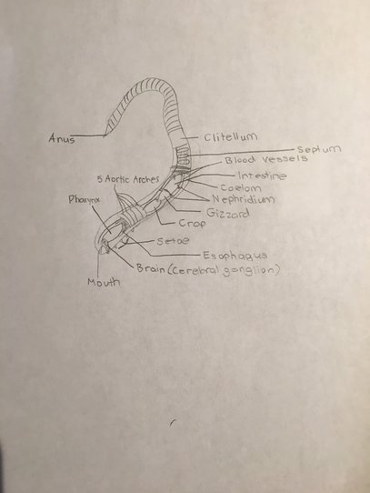
We started by taking our earthworm, placing it in the dissection tray, and identifying the dorsal and ventral sides. The dorsal side is usually more rounded, while the ventral side is flat. We turned our worm with its ventral side up and examined the external parts, such as the setae bristles and the clitellum, using a magnifying glass. Next, we used a scalpel to make an incision on its dorsal side from the head to the clitellum, and pinned the body walls open. Then we looked at the internal parts, the first being the five aortic arches, which are the five small tan bean shaped structures shown in photo 1. These function as the hearts. We also located the dorsal blood vessel, which is the long brown tube running down the whole worm. The pharynx, esophagus, crop, gizzard, and intestine were also examined. After removing the dorsal blood vessel, we could view the ventral nerve cord and the ganglion, as shown in photo 2. We discovered the nephridia were located on the right side of our worm- there is two in every segment. After drawing a quick sketch of our worm, shown to the right, we disposed of the parts and cleaned all tools and our work area. |
|
ANAYLSIS
1. What is the name of the pumping organs of an earthworm? The pumping organs are known as the five aortic arches. These pump blood through the dorsal and ventral vessels.
2. In the earthworm trace the parts of the digestive tract through which food passes. Earthworms are decomposers, which means they use their mucous covered pharynx to take in decaying matter. Other types filter out food particles from water into a large mucous bag. Earthworms specifically use their pharynx to transfer their food from the soil to their esophagus. It is then stored in their crop and broken down into smaller bits in their gizzard, before being absorbed into the intestine. Their nephridia excrete the metabolic wastes and any excess water afterwards out of the anus.
3. Which parts of the earthworm serve as its brain? How are these parts connected to the rest of the body? The earthworm's ganglion serve as the brain, located above the ventral nerve cord. This connects the ganglion to the rest of the body, as the nerve cord runs all the way from the mouth to the anus.
4. Which of the parts of the earthworm's body that you saw are included in the excretory system? All the parts of the digestive system, including the pharynx, esophagus, crop, and gizzard, as well as the nephridia. These work as a kidney to excrete wastes.
5. How can you find out whether an earthworm eats soil? You can really only tell whether an earthworm eats soil by dissecting it. If you slit open the pharynx, you will see a dark brown substance, like the stuff exhibited in photo 3 in the above slideshow. This is the absorbed soil that was taken in through the pharynx and was going to be transported to the esophagus, in order to extract food particles. This is the main sign that the earthworm eats soil, as that brown stuff inside of the pharynx is the soil itself.
6. Among the earthworm's structural adaptations are its setae. How do you think the earthworm's setae make it well adapted to its habitat? The setae help the earthworm hugely when it comes to movement, since they prevent slipping. However, setae also work together with the two groups of muscles called the hydrostatic skeleton- which contract to help the worm get shorter and fatter or vice versa. As a result, earthworms can burrow down into the soil, making them very adapted to their habitat since they can decompose dirt for food. They don't have to hunt in order to find food, they can easily obtain nutrients from the soil.
7. How is the earthworm's digestive system adapted for extracting relatively small amounts of food from large amounts of ingested soil? The earthworm has two very important parts of their digestive system called the crop and gizzard. The crop works to store food, and then the food moves to the gizzard, where it is crushed into smaller pieces before going to the intestine. As a result, the earthworm can take that ingested soil from the pharynx and once that soil gets to the gizzard, it can remove very small particles of food from very large amounts of soil.
8. Your dissection of the earthworm did not go beyond segment 32. What will you observe if you dissect the remainder of the worm to its posterior end? If we were to dissect past segment 32, also known as past the clitellum, we would see the intestine, and possibly some of the digested food that had been ground up in the gizzard.
9. What did each germ layer develop into in the earthworm? The earthworm is triploblastic, which means it has three germ layers: ectoderm, endoderm, and mesoderm. The ectoderm develops into the worms outer skin and its ventral cord and cerebral ganglion, which work as a nervous system to detect stimuli (such as statocysts for balance and ocelli for light). The endoderm develops into the lining of the digestive tract, so in the earthworm's case the lining of the pharynx, esophagus, crop, gizzard, and intestine. The mesoderm develops into the muscles and internal organs, so basically the earthworm's hydrostatic skeleton and organs such as the five Aortic arches, nephridia, digestive system, and blood vessels.
10. What is the function of the nephridium? How does it complete this function? The nephridium work like a kidney to remove metabolic wastes such as ammonia and excrete extra water. They complete this function by the wastes being diffused out of the earthworm's skin, while any food waste like excess soil exits out of the anus.
11. How is the sandworm similar and different to the earthworm? Both the sandworm and the earthworm are a part of the phylum Annelida, and have the same organ systems, such as the digestive and circulatory systems. However, for respiration, sandworms breathe through gills while earthworms use diffusion through their skin. Sandworms also have sharp pincers that capture prey, unlike earthworms. Both excrete wastes using their nephridia and their anus, and rely on water in order to survive. One of the biggest differences is that sandworms have appendages while earthworms have setae, for movement.
12. How is the leech designed to live as an ectoparasite? What are the similarities and differences between the leech and the earthworm? The leech is adapted so it feeds off of blood from a host, unlike other annelids. It attaches to the human using one sucker on its anterior to suck and another sucker on its posterior to stay anchored. This is different from earthworms since they take in food through a pharynx and ingest soil, and use setae to prevent slipping and move. This defines that leeches are parasitic, while earthworms are free-living. However, they are once again both part of the phylum Annelida, and need water to survive. They both have identical organ systems as well, not including the mouth structure. For excretion, they dispose of wastes using nephridia and their anus.
